43 simple microscope diagram with labels
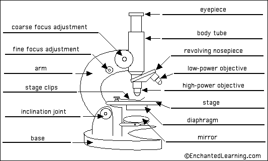
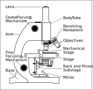
Microscope Parts and Functions A standard microscope has three, four, or five objective lenses that range in power from 4X to 100X. When focusing the microscope, be careful that the objective lens doesn't touch the slide, as it could break the slide and destroy the specimen. Specimen or slide: The specimen is the object being examined. Parts of the Microscope with Labeling (also Free Printouts) Parts of the Microscope with Labeling (also Free Printouts) By Editorial Team March 7, 2022 A microscope is one of the invaluable tools in the laboratory setting. It is used to observe things that cannot be seen by the naked eye. Table of Contents 1. Eyepiece 2. Body tube/Head 3. Turret/Nose piece 4. Objective lenses 5. Knobs (fine and coarse) 6.
Preface | Python Data Science Handbook - GitHub Pages Mar 26, 2013 · Whether you are reporting election results, forecasting stock returns, optimizing online ad clicks, identifying microorganisms in microscope photos, seeking new classes of astronomical objects, or working with data in any other field, the goal of this book is to give you the ability to ask and answer new questions about your chosen subject area.

Simple microscope diagram with labels
Microscope, Microscope Parts, Labeled Diagram, and Functions Microscope, Microscope Parts, Labeled Diagram, and Functions What is Microscope? A microscope is a laboratory instrument used to examine objects that are too small to be seen by the naked eye. It is derived from Ancient Greek words and composed of mikrós, "small" and skopeîn,"to look" or "see". Microscope Poster - Diagram with Labels | Teach Starter A poster containing a diagram with labels showing the key parts of a microscope. In Science it is important that students know how to use a variety of tools when conducting scientific experiments and inquiry. This poster focuses on the microscope and highlights its key parts. There are two print options available for this poster: Microscope With Labels clip art | Microscope parts, Scientific method ... Print a microscope diagram, microscope worksheet, or practice microscope quiz in order to learn all the parts of a microscope. Las partes de un microscopio tradicional pueden dividirse en doce partes (ver gráfico superior): 1) Ocular, 2) Tornillo macromético , 3) Tubo óptico , 4)
Simple microscope diagram with labels. Microscope Labeling - The Biology Corner 1) Start with scanning (the shortest objective) and only use the COARSE knob . Once it is focused… 2) Switch to low power (medium) and only use the COARSE knob . You may need to recenter your slide. Once it is focused.. 3) Switch to high power (long objective). Parts of a microscope with functions and labeled diagram - Microbe Notes Figure: Diagram of parts of a microscope There are three structural parts of the microscope i.e. head, base, and arm. Head - This is also known as the body. It carries the optical parts in the upper part of the microscope. Base - It acts as microscopes support. It also carries microscopic illuminators. Microscope Drawing Easy with Label - YouTube In this video I go over a microscope drawing that is easy with label. There is a blank copy at the end of the video to review on your own. A great way to s... Draw a neat (labelled) diagram for the formation of image in a simple ... Draw a neat (labelled) diagram for the formation of image in a simple microscope. Medium Solution Verified by Toppr Light from a light source (mirror) passes through a thin transparent object. A biconvex lens magnifies the size of the object to get an enlarged virtual image. The image is viewed from the other side. Video Explanation
Compound Microscope Parts, Functions, and Labeled Diagram The total magnification of a compound microscope is calculated by multiplying the objective lens magnification by the eyepiece magnification level. So, a compound microscope with a 10x eyepiece magnification looking through the 40x objective lens has a total magnification of 400x (10 x 40). Simple Microscope Definition, Magnification, Parts And Uses - BYJUS A simple microscope is a magnifying glass that has a double convex lens with a short focal length. Examples of this kind of instrument include the hand lens and reading lens. When an object is kept near the lens, then its principal focus with an image is produced, which is erect and bigger than the original object. 16 Parts of a Compound Microscope: Diagrams and Video Once you have an understanding of the parts of the microscope it will be much easier to navigate around and begin observing your specimen, which is the fun part! The 16 core parts of a compound microscope are: Head (Body) Arm Base Eyepiece Eyepiece tube Objective lenses Revolving Nosepiece (Turret) Rack stop Coarse adjustment knobs Labeling the Parts of the Microscope | Microscope World Resources Labeling the Parts of the Microscope This activity has been designed for use in homes and schools. Each microscope layout (both blank and the version with answers) are available as PDF downloads. You can view a more in-depth review of each part of the microscope here. Download the Label the Parts of the Microscope PDF printable version here.
Parts of a Microscope Labeling Activity - Storyboard That Create a poster that labels the parts of a microscope and includes descriptions of what each part does. Click "Start Assignment". Use a landscape poster layout (large or small). Search for a diagram of a microscope. Using arrows and textables label each part of the microscope and describe its function. Copy This Storyboard* More options Simple Microscope: Definition, Principle, Parts, And Uses In fact, most simple microscopes only have a 10x magnification power. The formula for calculating the magnifying power of a simple microscope is: M = 1 + D/F, where D is the least distance of distinct vision, and F is the focal length of the convex lens. The shorter the focal length of the lens, the higher the magnifying power of the microscope. Label the microscope — Science Learning Hub All microscopes share features in common. In this interactive, you can label the different parts of a microscope. Use this with the Microscope parts activity to help students identify and label the main parts of a microscope and then describe their functions. Drag and drop the text labels onto the microscope diagram. Simple Microscope - Definition, Types, Working Principle & Formula A simple microscope consists of a convex lens of a short focal length. The below figure shows the ray diagram which subsequently forms the image of an object (or we can say a source of light). (Image will be Updated soon) F is the focal length of the lens. An object is placed between the focal length and the centre of the curvature.
29 best Anatomy and Physiology images on Pinterest | Physiology, Anatomy and Anatomy reference
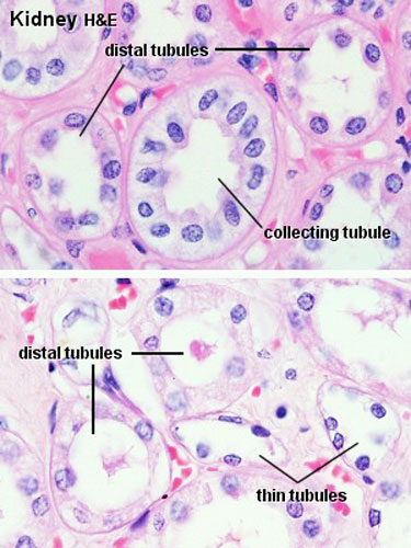
Simple Squamous Epithelium under a Microscope with a Labeled Diagram ... From the lung parenchyma labeled diagram, you might identify the following structures - Simple squamous epithelium lining of the lung alveoli (within the parenchyma), A connective tissue basement membrane beneath the simple squamous epithelium lining, The lumen of the lung alveoli, and The cytoplasm of the simple squamous epithelium cells.

Pin on Diagrams/Charts/Maps
Microscope Labeling - The Biology Corner Students label the parts of the microscope in this photo of a basic laboratory light microscope. Can be used for practice or as a quiz. ... The type of microscope used in most science classes is the _____ microscope. 18. You should carry the microscope by the _____ and the _____. 19. The objectives are attached to what part of the microscope ...
Plant cell Structure: Plant cell parts, Organelles and their functions and Diagram
Fluorescence Resonance Energy Transfer (FRET) Microscopy Presented in Figure 3 is a Jablonski diagram illustrating the coupled transitions involved between the donor emission and acceptor absorbance in fluorescence resonance energy transfer. Absorption and emission transitions are represented by straight vertical arrows (green and red, respectively), while vibrational relaxation is indicated by wavy ...

Cell Biology ~ Pass. Science. Solutions.
Simple Microscope - Parts, Functions, Diagram and Labelling Simple Microscope - Parts, Functions, Diagram and Labelling By Editorial Team March 7, 2022 A microscope is one of the commonly used equipment in a laboratory setting. A microscope is an optical instrument used to magnify an image of a tiny object; objects that are not visible to the human eyes. Table of Contents

Clipart Panda - Free Clipart Images
Compound Microscope Parts - Labeled Diagram and their Functions The eyepiece (or ocular lens) is the lens part at the top of a microscope that the viewer looks through. The standard eyepiece has a magnification of 10x. You may exchange with an optional eyepiece ranging from 5x - 30x. [In this figure] The structure inside an eyepiece. The current design of the eyepiece is no longer a single convex lens.
Foundations - Histology Epithelia and Skin - Embryology
Fluorescence - Wikipedia Fluorescence is the emission of light by a substance that has absorbed light or other electromagnetic radiation.It is a form of luminescence.In most cases, the emitted light has a longer wavelength, and therefore a lower photon energy, than the absorbed radiation.

The Microscope: Create a Labelled Diagram | Teaching Resources
Label the Microscope Diagram | Download Scientific Diagram - ResearchGate Download scientific diagram | Label the Microscope Diagram from publication: Laboratory Exercises in Microbiology: Discovering the Unseen World through Hands-on Investigation | Microbiology ...
Microscope labelling 11 - Teaching resources
Microscope labeled diagram - slideshare.net The Microscope Image courtesy of: Microscopehelp.com Basic rules to using the microscope 1. You should always carry a microscope with two hands, one on the arm and the other under the base. 2. You should always start on the lowest power objective lens and should always leave the microscope on the low power lens when you finish using it. 3.

Microscope Unlabelled Diagram - Micropedia
Stages of transcription - Khan Academy In the microscope image shown here, a gene is being transcribed by many RNA polymerases at once. The RNA chains are shortest near the beginning of the gene, and they become longer as the polymerases move towards the end of the gene. This pattern creates a kind of wedge-shaped structure made by the RNA transcripts fanning out from the DNA of the ...
1.1 Labelling Microscope - Labelled diagram
GCSE Science: Required practical activities - AQA Using a light microscope to observe, draw and label cells in an onion skin . Materials . In addition to access to general laboratory equipment, each student needs: • a small piece of onion • a knife or scalpel • a white tile • forceps • a microscope slide • a coverslip • a microscope • iodine solution in a dropping bottle.

Simple Cuboidal
Labelled Diagram of Compound Microscope - Biology Discussion The below mentioned article provides a labelled diagram of compound microscope. Part # 1. The Stand: The stand is made up of a heavy foot which carries a curved inclinable limb or arm bearing the body tube. The foot is generally horse shoe-shaped structure (Fig. 2) which rests on table top or any other surface on which the microscope in kept.

Labelled Diagram Of A Tick - Top Label Maker
Parts of a Simple Microscope - Labeled (with diagrams) The metal stand of a simple microscope has two primary components - base plate and vertical rod. The metal stand supports the entire parts of the microscope. It also provides stability to other parts. Stage It is a plate made of metal and rectangular in shape. It has a hole in the center which allows the light from the below to pass through.

11 Best Images of Cell Labeling Worksheet Answers - Cell Cycle and Mitosis Worksheet Answers ...
Microscope Label Worksheets & Teaching Resources | TpT Label the Microscope by Crista Tiboldo 19 FREE PDF (178.64 KB) Worksheet identifying the parts of the compound light microscope. Answer key: 1. Body tube 2. Revolving nosepiece 3. Low power objective 4. Medium power objective 5. High power objective 6. Stage clips 7. Diaphragm 8. Light source 9. Eyepiece 10. Arm 11. Stage 12.
Biology 521 Resources
Microscope Types (with labeled diagrams) and Functions Simple microscope labeled diagram Simple microscope functions It is used in industrial applications like: Watchmakers to assemble watches Cloth industry to count the number of threads or fibers in a cloth Jewelers to examine the finer parts of jewelry Miniature artists to examine and build their work Also used to inspect finer details on products
Microscope With Labels clip art (111146) Free SVG Download / 4 Vector
The Parts of a Microscope (Labeled) Printable - TeacherVision The Parts of a Microscope (Labeled) Printable. Download. Add to Favorites. Share. This diagram labels and explains the function of each part of a microscope. Use this printable as a handout or transparency to help prepare students for working with laboratory equipment.
Post a Comment for "43 simple microscope diagram with labels"