39 fluorescent labels and light microscopy
Different Ways to Add Fluorescent Labels - Thermo Fisher Scientific Labeling various targets with separate fluorescent colors allows you to visualize different structures or proteins within a cell in the same experiment. Ways to fluorescently label your target include fluorescent dyes, immunolabeling, and fluorescent fusion proteins —all of which can provide a means to selectively mark structures and proteins ... Fluorescent Labeling - What You Should Know - PromoCell Definition Fluorescent labeling is the process of binding fluorescent dyes to functional groups contained in biomolecules so that they can be visualized by fluorescence imaging (nature.com). The availability of new fluorophores has dramatically changed the possibilities for the sensitive detection of biomolecules and the analysis of their interactions. Improved fluorescent dyes are now ...
Recent Advances in Fluorescent Labeling Techniques for ... by T Suzuki · 2007 · Cited by 106 — The techniques of fluorescent labeling of molecules are divided broadly into two categories: the conventional fluorescent staining and the molecular tagging by ...

Fluorescent labels and light microscopy
Fluorescence Microscopy - an overview | ScienceDirect Topics M. Heilemann, in Comprehensive Biophysics, 2012 Abstract. Fluorescence microscopy is a valuable toolbox to study cellular structures and dynamics spanning scales from the single molecule to the live animal. The spatial resolution that can be achieved with any light-based microscopy is however limited to about 200 nm in the imaging plane and >500 nm along the optical axis. Integrating High Resolution Light Microscopy and Real Time ... by TA Hasling · 2006 · Cited by 3 — The classic fluorescent techniques (which include epifluorescence and confocal) allow researchers to selectively observe labeled structures with ... Fluorescence microscope - Wikipedia The majority of fluorescence microscopes, especially those used in the life sciences, are of the epifluorescence design shown in the diagram.Light of the excitation wavelength illuminates the specimen through the objective lens. The fluorescence emitted by the specimen is focused to the detector by the same objective that is used for the excitation which for greater resolution will need ...
Fluorescent labels and light microscopy. Fluorescent tag - Wikipedia Fluorescent tag. S. cerevisiae septins revealed with fluorescent microscopy utilizing fluorescent labeling. In molecular biology and biotechnology, a fluorescent tag, also known as a fluorescent label or fluorescent probe, is a molecule that is attached chemically to aid in the detection of a biomolecule such as a protein, antibody, or amino acid. Introduction to Fluorescence Microscopy | Nikon's MicroscopyU Introduction to Fluorescence Microscopy. The absorption and subsequent re-radiation of light by organic and inorganic specimens is typically the result of well-established physical phenomena described as being either fluorescence or phosphorescence. The emission of light through the fluorescence process is nearly simultaneous with the ... Advanced Fluorescence Microscopy Techniques—FRAP, FLIP, FLAP ... Apr 02, 2012 · 1. Introduction. FRAP, FLIP, FLAP, FRET, and FLIM are fluorescence microscopy techniques that in some way take advantage of particular aspects of the fluorescence process by which fluorochromes are excited and emit fluorescent light, are damaged during repetitive excitation, or undergo non-radiative decay prior to light emission. Label-free prediction of three-dimensional fluorescence images from ... Although fluorescence microscopy can resolve subcellular structure in living cells, it is expensive, is slow, and can damage cells. ... phenotypes can be detected via expressed fluorescent labels ...
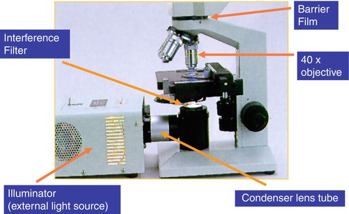
Basics of FRET Microscopy | Nikon’s MicroscopyU The first fluorescent protein biosensor was a calcium indicator named cameleon, constructed by sandwiching the protein calmodulin and the calcium calmodulin-binding domain of myosin light chain kinase (M13 domain) between enhanced blue and green fluorescent proteins (EBFP and EGFP). In the presence of increasing levels of intracellular calcium ... Researchers demonstrate label-free super-resolution microscopy A newly developed sub-diffraction-limit microscopy approach doesn't require fluorescent labels. The video shows the process of the data evaluation algorithm, retrieving the positions and sizes ... New fluorescent label provides a clearer picture of how DNA ... Unlike traditional fluorescence microscopy, which uses labels that glow constantly, this approach involves switching on only a subset of the labels at each moment. Fluorescent Microscopy - SERC A fluorescence microscope is much the same as a conventional light microscope with added features to enhance its capabilities. The conventional microscope uses visible light (400-700 nanometers) to illuminate and produce a magnified image of a sample. A fluorescence microscope, on the other hand, uses a much higher intensity light source which ...
Fluorescence Microscopy - Explanation and Labelled Images A fluorescence microscope works by combining the magnifying properties of the light microscope with fluorescence emitting properties of compounds. Fluorescence microscopy uses a high-intensity light source that excites a fluorescent molecule called a fluorophore in the sample observed. ... and by doing so, highlight (or "label") the nuclei ... A quick guide to light microscopy in cell biology FLUORESCENCE MICROSCOPY · by K Thorn · 2016 · Cited by 143 — Most molecules in the cell are not very fluorescent, so fluorescent labels to be imaged are typically introduced by the ... Label-free prediction of three-dimensional fluorescence images from ... Although fluorescence microscopy can resolve subcellular structure in living cells, it is expensive, is slow, and can damage cells. We present a label-free method for predicting three-dimensional fluorescence directly from transmitted-light images and demonstrate that it can be used to generate multi-structure, integrated images. Fluorescence Microscopy & Cell Imaging | Research | UNM Cancer Center Imaging. The Fluorescence Microscopy and Cell Imaging Shared Resource aids basic and physician researchers to image samples and publish high profile articles that: Elucidate cell and molecular mechanisms of cancer, immunologic, infectious, metabolic, neurologic and vascular diseases. Evaluate therapeutic efficacy in cells and patient samples.
Read more about Superresolution Microscopy
Introduction to Fluorescent Proteins | Nikon's MicroscopyU The brightness and fluorescence emission spectrum of enhanced yellow fluorescent protein combine to make this probe an excellent candidate for multicolor imaging experiments in fluorescence microscopy. Enhanced yellow fluorescent protein is also useful for energy transfer experiments when paired with enhanced cyan fluorescent protein (ECFP) or ...

Correlative light and electron microscopy fundamentals
Labeling the ER for Light and Fluorescence Microscopy Most of them are not 100% specific for the ER membrane and may label other organelles at varying concentrations and incubation times. ... C., Wang, P., Kriechbaumer, V. (2018). Labeling the ER for Light and Fluorescence Microscopy. In: Hawes, C., Kriechbaumer, V. (eds) The Plant Endoplasmic Reticulum . Methods in Molecular Biology, vol 1691 ...
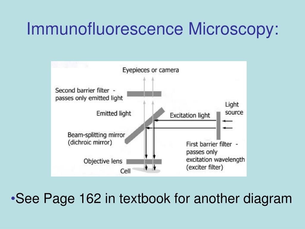
PPT - Immunofluorescence Microscopy PowerPoint Presentation, free download - ID:6715484
Labeling the ER for Light and Fluorescence Microscopy by C Hawes · 2018 · Cited by 3 — The ER is a highly dynamic network of tubules and membrane sheets. Hence imaging this organelle in its native and mobile state is of great importance.

Science Visualized • LIGHT SHEET FLUORESCENCE MICROSCOPY Light sheet...
Fluorescence microscopy: established and emerging methods, experimental ... Numerous practical strategies to enhance fluorescence microscopy experiments are reviewed. The use of instrumentation such as light traps, cameras, objectives, improved fluorescent labels, and image filtration routines applicable to low light level experiments are discussed.

Label-free prediction of three-dimensional fluorescence images from transmitted light microscopy ...
Fluorescent Labelling - an overview | ScienceDirect Topics The light source used in the fluorescence microscope is generally a high-brightness light source, such as a xenon or mercury lamp (Aswani et al., 2012). These two types of arc lamps were selected based on the measurement object.

Confocal laser scanning microscopy of plant cells labeled with various... | Download Scientific ...
Dots, Probes and Proteins: Fluorescent Labels for Microscopy and Imaging GFP now comes in 'flavors' including cyan, yellow and blue. Fluorescent proteins are useful for studying live cells and can be used as 'reporters' for studying gene expression. Using genetically modified plasmid and/or viral DNA, the target cells can be transfected with the plasmid which encodes both the fluorescent protein and a gene ...
Biomedical engineering department celebrates five years - GW Hatchet
Novel Fluorescent Label Shines a Light on DNA Structure in Cancer Cells March 7, 2022. 0. Researchers have developed a new fluorescent label that gives a clearer picture of how DNA architecture is disrupted in cancer cells. The findings could improve cancer diagnoses ...
Microscopy Stock Images, Royalty-Free Images & Vectors | Shutterstock
Super resolution fluorescence microscopy - PMC Among the various microscopy techniques, fluorescence microscopy is one of the most widely used because of its two principal advantages: Specific cellular components may be observed through molecule-specific labeling, and light microscopy allows the observation of structures inside a live sample in real time.

What is Correlative Light and Electron Microscopy?
Fluorescent labeling of abundant reactive entities (FLARE) for ... - Nature Fluorescence microscopy is a vital tool in biomedical research but faces considerable challenges in achieving uniform or bright labeling. For instance, fluorescent proteins are limited to model ...
FLUORESCENCE MICROSCOPY/CELL BIOLOGY: The planar truth about light-sheet microscopy | Laser ...
Multispectral intravital microscopy for simultaneous bright ... - Springer Conventional light microscopes do not allow for simultaneous bright-field and fluorescent imaging. Moreover, in conventional microscopes, only one type of fluorescent label can be observed. This study introduces multispectral intravital video microscopy, which combines bright-field and fluorescence microscopy in a standard light microscope.
Fluorescent Microscopy and Fluorescent Labelling for Malaria Diagnosis | SpringerLink
Quantum dots versus organic dyes as fluorescent labels ... Aug 28, 2008 · In contrast, blinking may be exploited for superresolution microscopy by analyzing the intermittent fluorescence to allow identification of the light emitted by each individual label and to ...
PolySciTech® - Flamma Fluors - Fluorescent Probes and Dyes
Fluorescence Imaging - Teledyne Photometrics By targeting these fluorescent labels, researchers can select what they want to see. This is demonstrated in Fig.3, ... Two-photon Fluorescence Light Microscopy. Macmillan Publishing Group. Schermelleh, L., Heinztmann, R., and Leonardt, H. (2010). A Guide to Super-Resolution Fluorescence Microscopy. The Journal of Cell Biology 190 (2): 165-175.
ZEISS Microscopy Online Campus | Introduction to Photoactivated Localization Microscopy (PALM)
Imaging Flies by Fluorescence Microscopy: Principles, Technologies, and ... The development of fluorescent labels and powerful imaging technologies in the last two decades has revolutionized the field of fluorescence microscopy, which is now widely used in diverse scientific fields from biology to biomedical and materials science. ... has brought about the era of fluorescence light microscopy. The first fluorescence ...

Label-free prediction of three-dimensional fluorescence images from transmitted-light microscopy ...
Light sheet fluorescence microscopy breakthrough - 2018 - Wiley ... Light-sheet fluorescence microscopy in single- and two-photon modes has emerged as a powerful wide-field, low-photodamage technique for fast volumetric imaging of biological samples. However, as Dholakia explain in Optics Letters, he and colleagues wanted to extend this imaging modality to the three-photon regime, enhancing its penetration ...

Development a flexible light‐sheet fluorescence microscope for high‐speed 3D imaging of calcium ...
Label-free prediction of three-dimensional fluorescence images from ... Fluorescence microscopy can resolve subcellular structure in living cells, but is expensive, slow, and toxic. Here, we present a label-free method for predicting 3D fluorescence directly from transmitted light images and demonstrate its use to generate multi-structure, integrated images.

Label-free prediction of three-dimensional fluorescence images from transmitted-light microscopy ...
Fluorescence Microscopy vs. Light Microscopy - Medical News This light is in the 400-700 nm range, whereas fluorescence microscopy uses light with much higher intensity. The usefulness of traditional light microscopy is hampered by the fact that it uses ...
Post a Comment for "39 fluorescent labels and light microscopy"